


Publications

Tendotonometry allows noninvasive in vivo biomechanical evaluation of the patellar tendon in sedated dogs in a study assessing intrauser variability
Authors: Kayla C. Caturay 1, Nicole S. Amato 1, Samuel D. Stewart 2, Chand Khanna 2, Heather Wilson-Robles 2, David M. Schmidt 1, Mitchell T. Roske 1, Kurt Schulz 2, 3
Affiliations:
- Massachusetts Veterinary Referral Hospital, Ethos Veterinary Health, Woburn, MA, USA
- Ethos Discovery, San Diego, CA, USA
- Ethos Peak Veterinary Referral Center, Ethos Veterinary Health, Williston, VT, USA
Journal: American Journal of Veterinary Research - August 2026 (DOI: 10.2460/ajvr.26.05.0228)
-
Field & Applications:
- Animal studies
- Medical
- Orthopedics
- Muscle symmetry
- Reliability
Objective: To describe the use and determine the feasibility and user reliability of a commercially available digital palpation device, a myotonometer, on the canine patellar tendon (PT).
Methods: In this prospective clinical feasibility study, client-owned dogs with no history of pelvic limb lameness were eligible for study enrollment from September through December of 2025 and included if their stifles were considered normal based upon physical examination and radiographic findings. Dogs were sedated, and a myotonometer was used to measure tone, stiffness, elasticity, relaxation, and creep of the PT. For each PT, a single user obtained 2 measurement series consisting of 5 individual measurements.
Results: 20 dogs were enrolled, and 400 total tendotonometric measurements were collected. No adverse events were observed. There were no statistically significant differences for any of the 5 biomechanical parameters between series within each side nor between left and right groups. There was good to excellent intraobserver repeatability between series and moderate to poor repeatability between sides.
Conclusions: This clinical study demonstrated feasible myotonometer use as a novel method to obtain biomechanical data for the canine PT in a cohort of sedated dogs with normal stifles.
Clinical Relevance: This study introduces tendotonometry into the clinical setting as a novel modality for in vivo evaluation of orthopedic soft tissues using the PT as a test case. Beyond the canine PT, future tendotonometry studies may allow for the generation of noninvasive, rapid, and objective biomechanical data in tendon repair and healing, soft tissue reconstruction for orthopedic conditions, and treatment response in neuromuscular disorders.
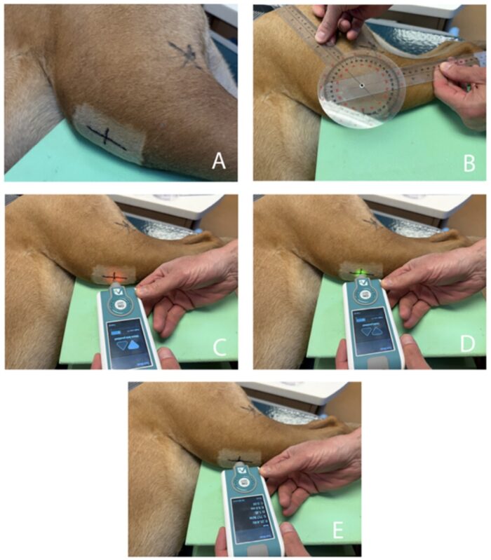
Figure 1. Images of a sedated dog with relaxed limb position undergoing left patellar tendon (PT) evaluation with a commercially available myotonometer during a study conducted from September through December of 2025 that included 20 dogs and 400 tendotonometric measurements to determine the feasibility and user reliability of the device for noninvasive in vivo biomechanical evaluation of PTs in dogs. A—A proximodistal line is drawn immediately distal to the patella to delineate the PT. A mediolateral line is drawn to bisect the length of the PT. The intersection of the 2 lines demarcates the middle of the PT. B—A goniometer is used to note the stifle angle. C—The illuminated red light indicates a measurement series is ready to be obtained. The myotonometer probe is positioned perpendicular over the center of the object of interest (intersection of the 2 lines). D—Illumination of the green light indicates an appropriate probe position within a range of 3 mm to obtain measurements. E—Upon completion of a measurement series, the calculated averages for tone, stiffness, and decrement populate, and the light is no longer illuminated.
Keywords: patellar tendon, dog, tendotonometry, stiffness, biomechanical
This clinical feasibility study demonstrates that tendotonometry can provide objective biomechanical data of the canine PT in vivo in sedated dogs weighing 13 to 52 kg using a commercial myotonometer with no known user challenges nor adverse events. The results of this study may be used as the basis for ongoing studies to define both intra- and interuser variability in a similar unaffected cohort of dogs, to evaluate PT characteristics between sound and lame dogs with PT abnormalities, and to compare awake versus sedated evaluation. Beyond the canine PT, the use of tendotonometry may allow for the generation of noninvasive, rapid, and objective biomechanical data in tendon repair and healing, soft tissue reconstruction for orthopedic conditions, and treatment response in acquired and congenital neuromuscular disorders. In the long term, we will continue to ask if and how tendotonometry may serve as a diagnostic tool to reliably assess other musculoskeletal structures in veterinary patients and what potential clinical applications it may have.