


Publications

Myotonometry and Muscle Force in Patients with Surgically Treated Tibial Pilon Fracture: A Cross-Sectional Study
Authors: Andrei-Daniel Bolovan 1, 2, Gheorghe-Bogdan Hogea 3, 4, 5, Elena-Constanta Amaricai 2, Alexandra-Roxana Tapardea 1, Ahmed Abu-Awwad 3, 4, 5, Liliana Catan 2
Affiliations:
- Doctoral School, “Victor Babes” University of Medicine and Pharmacy, 300041 Timisoara, Romania
- Research Center for Assessment of Human Motion, Functionality and Disability, Department of Rehabilitation, Physical Medicine and Rheumatology, “Victor Babes” University of Medicine and Pharmacy, 300041 Timisoara, Romania
- Department of Orthopedics and Traumatology, “Victor Babes” University of Medicine and Pharmacy, 300041 Timisoara, Romania
- “Pius Brinzeu” Emergency Clinical County Hospital, Bld. Liviu Rebreanu, No. 156, 300723 Timisoara, Romania
- Research Center Teodor Sora, Department of Orthopedics II, “Victor Babes” University of Medicine and Pharmacy, Eftimie Murgu Square, No. 2, 300041 Timisoara, Romania
Journal: Journal of Functional Morphology and Kinesiology - January 2026, Volume 11, Issue 1, Article no. 21 (DOI: 10.3390/jfmk11010021)
-
Field & Applications:
- Medical
- Orthopedics
- Recovery from injury
- Musculoskeletal rehabilitation
- Reliability
Background: Tibial pilon fractures are, in most cases, complex injuries caused by high-energy trauma. This type of fracture requires surgical stabilization and immobilization that impairs ankle function by reducing range of motion, muscle strength, and affecting the mechanical properties of the muscles.
Methods: We evaluated 22 patients who required surgery for tibial pilon fractures and 22 age-matched healthy controls. Dynamometry assessed the isometric strength of the dorsiflexors and plantar flexors. Myotonometry of the tibialis anterior, peroneus longus, and medial and lateral gastrocnemius muscles analyzed the muscle tone, biomechanical (stiffness and decrement), and viscoelastic properties (mechanical stress relaxation and ratio of relaxation time to deformation time (creep).
Results: Compared to the control group, the patients had significantly decreased isometric strength in both the dorsal flexors and plantar flexors on the affected side. Myotonometric measurements did not reveal significant differences in the tibialis anterior and peroneus longus muscles. Both medial and lateral gastrocnemius muscles exhibited significantly increased frequency and stiffness, and significantly decreased relaxation and creep in patients when compared to the control group.
Conclusions: When compared to healthy controls, patients with surgically treated unilateral pilon fracture had a decreased isometric muscle force of ankle dorsiflexors and plantar flexors of both affected and non-affected lower limbs. Myotonometry indicated increased frequency and stiffness, along with decreased values of viscoelastic parameters (stress relaxation time and creep) in the medial and lateral gastrocnemius muscles on both sides.
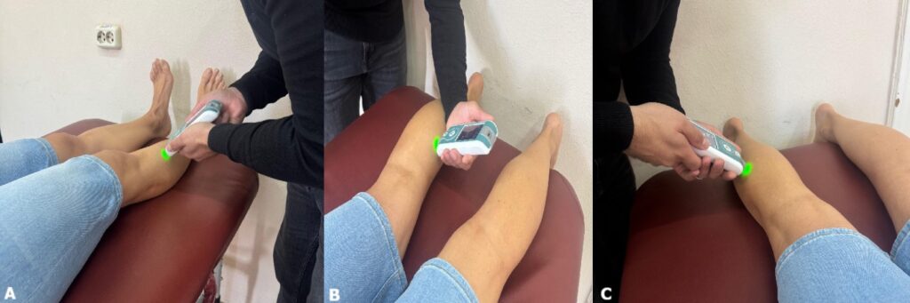
Figure 2. Myotonometric assessment points and participant positions for the tibialis anterior (A), medial gastrocnemius (B), and lateral gastrocnemius (C) muscles.
Keywords: tibial pilon fracture, myotonometry, stiffness, muscle strength, dynamometry
Patients with surgically treated unilateral tibial pilon fractures showed reduced isometric muscle strength in ankle dorsiflexors and plantar flexors on both the affected and unaffected limbs, compared to healthy controls. Myotonometry showed increased frequency and stiffness, along with decreased viscoelastic parameters (stress relaxation time and creep) in the medial and lateral gastrocnemius muscles on both sides.